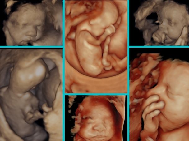
Technological advancement has revolutionized how we experience pregnancy, particularly regarding baby ultrasounds. These imaging procedures allow expectant parents to glimpse their little ones before birth, creating a special connection and a sense of wonder. But have you ever wondered how ultrasounds work? This blog will delve into the science behind baby ultrasounds, explicitly focusing on 3D/4D ultrasound imaging. From understanding sound waves to creating detailed images, let’s explore the fascinating world of baby ultrasounds.
Imagine sitting in a dimly lit room filled with anticipation and excitement. The soft sound of a heartbeat fills the air as you watch the screen before you. Suddenly, a clear image appears, revealing the tiny features of your little one—the button nose, the delicate fingers, and the gentle movements.
It’s a moment of pure wonder and awe as you witness the miracle of life unfolding before your eyes. This magical experience is made possible by the science behind baby ultrasounds, specifically the remarkable 3D/4D ultrasound imaging technology. In this blog, we will explore the intricacies of this process and unravel the mysteries of how sound waves create beautiful images of your little one inside the womb.
Understanding Sound Waves:
At the heart of baby ultrasounds lies the concept of sound waves. In the case of ultrasounds, high-frequency sound waves capture images of the developing baby in the womb. These sound waves are produced by a transducer, a handheld device that emits and receives the waves.
The Transducer: The Key to Imaging:
The transducer is a vital component of the ultrasound machine. It consists of a probe that emits sound waves and a receiver that captures the waves bouncing back. The investigation is placed on the expectant mother’s abdomen, and a conductive gel is applied to ensure proper contact and sound wave transmission.
Sound Wave Reflection and Echoes:
When the sound waves emitted by the transducer encounter different tissues and structures inside the mother’s body, they bounce back, creating echoes. These echoes are received by the transducer’s receiver and converted into electrical signals. The timing and strength of these echoes provide information about the location and characteristics of the tissues and structures.
Image Formation: From Echoes to Visuals:
The ultrasound machine’s computer processes the electrical signals produced by the echoes. Using sophisticated algorithms, the computer visually represents the echoes received. This results in detailed images that depict the baby’s internal structures, including organs, bones, and movements.
2D Ultrasound: A Classic Approach:
The most common type of baby ultrasound is the 2D ultrasound. It produces flat, two-dimensional images that provide valuable information about the baby’s growth, position, and development. These images are created by capturing echoes from multiple angles and piecing them together to form a coherent picture.
3D Ultrasound: Adding Depth and Dimension:
With technological advancements, 3D ultrasound imaging has taken the experience to a new level. 3D ultrasounds use the same principles as 2D ultrasounds but incorporate additional information to create three-dimensional images. By capturing echoes from multiple angles, the computer generates a lifelike representation of the baby, allowing expectant parents to see their little one’s facial features, limbs, and body contours
4D Ultrasound: The Fourth Dimension of Time:
If three dimensions are not enough, there’s the marvel of 4D ultrasound. 4D ultrasound takes the three-dimensional images a step further by adding the dimension of time. This means that expectant parents can witness the real-time movements of their babies. They can see the baby yawning, stretching, and smiling, providing a unique and emotional connection.
The Safety of Ultrasound Imaging:
One common concern regarding 6 week ultrasound is the safety of both the mother and the baby. Numerous studies have been conducted to assess the safety of ultrasound imaging, and the consensus is that it is considered safe when used as directed by healthcare professionals. The sound waves in ultrasounds have low energy levels, and the exposure time is typically short. However, following medical guidelines and avoiding unnecessary and prolonged exposure is essential.
The Joy and Bonding of Baby Ultrasounds:
Beyond science and technology, baby 6 week ultrasound pictures offer a profound emotional experience for expectant parents. They provide a unique opportunity to bond with the baby before birth, fostering a sense of connection and love. Seeing the baby’s face, movements, and gestures on the ultrasound screen can create memories that last a lifetime.
Conclusion:
Baby ultrasounds, including baby 3D/4D ultrasound imaging, are a remarkable application of sound wave technology. From producing sound waves to forming detailed images, the science behind baby ultrasounds is awe-inspiring. These imaging procedures allow expectant parents to witness their little one’s incredible development journey and foster a deep emotional connection. As technology advances, baby’s ultrasound will continue to bring joy, wonder, and cherished memories to countless families worldwide.


